Common Applications
| Confocal: | Confocal imaging is a fluorescence microscopy technique that optically sections the specimens preventing out of focus light from reaching the detector. This yields clear high contrast images, and together with the ability to acquire images sequentially at multiple positions the entire sample can be reconstructed in 3D. |
| Live Cell Imaging: | Many biological processes are dynamic events and as such observing living cells is crucial to unravelling and understanding the mechanisms behind them. This requires special consideration when imaging. Firstly, the environmental conditions must be controllable and stable to ensure the cell remain healthy. Secondly, care needs to be taken that the act of imaging has as little impact as possible on the event being observed. |
| FRAP: | Fluorescence Recovery After Photobleaching is used to gain insight into the dynamics or both diffusion and binding rates within a sample. Photobleaching is an irreversible process by which a fluorophore loses the ability to absorb and emit light. By deliberately inducing photobleaching within a restricted region of the sample observing the recovery within this region over time is affected by the rate at which unbleached molecules entre this region and bleached molecules are moved. The light sheet mode available on this instrument allows to study thicker samples than (<2mm) what an ordinary confocal microscope could image |
| Photo-activation |
Photoactivation is a technique of photomanipulation used to investigate intracellular protein movement. A laser is used to cause a structural change to a population of molecules like fluorophores. These photoactivated molecules can then be tracked to assess their movements. |
| Tissue Clearing |
Tissue clearing is a preparation technique with the aim of reducing the inherently scattering nature of tissue. There are several means by which this is achieved but all enable clearer and deeper penetration into the sample using light microscopy. |
| Digital Lightsheet Microscopy |
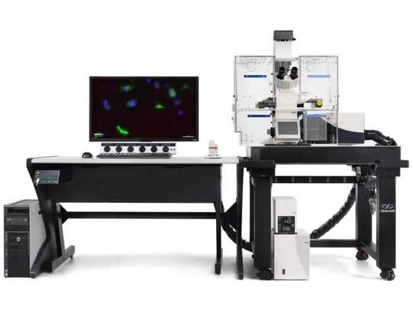
The Leica TCS SP8 DLS is based on an ordinary confocal microscope. To convert such a system into a digital light sheet microscope, small mirrors are positioned just outside the observed field, at the position of the focal plane and on each side of the specimen, to make the illumination beam pass through the sample perpendicular to the optical axis. The lateral scanning modes is used to scan the illumination line in the perpendicular direction, creating the required two-dimensional light sheet. The deflecting mirrors are mechanically connected to the observation lens to ensure the illuminated z-position is always in the focus of the image-generating optics. The image is then projected onto a video camera that converts the intensities into a digital image. |
| Colocalisation |
Light microscopy lends itself very well to labelling multiple structures within a sample due to the ability to separate these spatially overlapping signals by the wavelength of light they emit. Colocalisation is the study of how the distribution of one probe relates to that of another within the same sample. |


