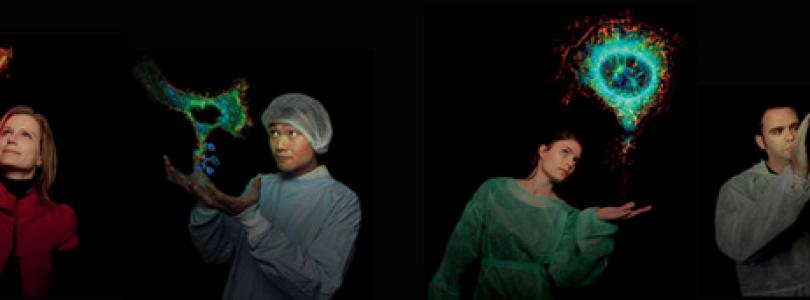
The Katharina Gaus Light Microscopy Facility was established in 2009 with the aim to build a fluorescence microscopy facility of the highest international standard. Currently, the facility contains over 40 of the latest advanced fluorescence microscopes and associated spectroscopy equipment which are supported by a group of specialist staff.
The core mission of the facility is to
- Conduct excellent training in specimen preparation, acquisition, and analysis of fluorescence microscopy data
- Provide high-end research support to scientists dedicated to biomedical research
- Provide access to expertise, and enable usage of state-of-the-art imaging
- Conduct collaborative research in technique development and analysis solutions
- Develops frontier technologies in bioengineering and bionanotechnology
- To lead Australia in the establishment and testing of new microscopes and imaging technologies
Falling within the UNSW Mark Wainwright Analytical Centre (MWAC), the Katharina Gaus Light Microscopy Facility has become a world-class imaging facility that enables innovative, cross-disciplinary research at the forefront of fundamental biomolecular sciences. The BMIF is particularly strong in cell and single-molecule imaging.
Our microscopes enable:
- 2-photon and Intravital Microscopy
- Biological / Soft Material Atomic Force Microscopy (AFM)
- Confocal Microscopy - Confocal Laser Scanning Microscopy (CLSM)
- Airy scan microscopy
- Epifluorescence for live cell imaging
- Fluorescence Correlation Spectroscopy (FCS)
- Fluorometer and Lifetime Spectroscopy
- Spinning Disc Microscopy
- Stimulated Emission Depletion (STED) Microscopy
- Stochastic Optical Reconstruction Microscopy (STORM)
- Superresolution Fluorescence Microscopy including photo-activation localisation microscopy (PALM)
- Time-Resolved Single-Molecule Imaging including Fluorescence Lifetime Imaging Microscopy (FLIM)
- Total Internal Reflection Fluorescence (TIRF) Microscopy
- Structured Illumination Microscopy (SIM)
- Apotome - Structured Illumination
- Gaussian and Lattice Lightsheet microscopy imaging (LSFM)
- Slide scanning (Brightfield, darkfield, phase contrast, polarized and fluorescence)
- Multiplex fluorescent microscopy imaging and spectral unmixing
- Second-Harmonic Generation (SHG) microscopy
- Ptychography - Quantitative Phase Imaging (QPI)


